
Online Database of Chemicals from Around the World
| Nanjing Wali Chemical Technology Co., Ltd. | China | |||
|---|---|---|---|---|
 | www.njwali.com | |||
 | 18005141382 | |||
 | 18005141382@163.com | |||
 | QQ Chat | |||
 | WeChat: 18005141382 | |||
| Chemical manufacturer since 2008 | ||||
| chemBlink Standard supplier since 2026 | ||||
| Classification | Organic raw materials >> Aldehyde |
|---|---|
| Name | 9-Cis-Retinal |
| Synonyms | (2E,4E,6Z,8E)-3,7-dimethyl-9-(2,6,6-trimethylcyclohexen-1-yl)nona-2,4,6,8-tetraenal |
| Molecular Structure | 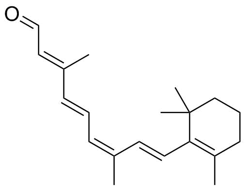 |
| Molecular Formula | C20H28O |
| Molecular Weight | 284.44 |
| CAS Registry Number | 514-85-2 |
| EC Number | 208-188-8 |
| SMILES | CC1=C(C(CCC1)(C)C)/C=C/C(=CC=CC(=CC=O)C)/C |
| Density | 0.9±0.1 g/cm3 Calc.* |
|---|---|
| Melting point | 56 - 58 °C (Expl.) |
| Boiling point | 421.4±14.0 °C 760 mmHg (Calc.)* |
| Flash point | 205.4±12.4 °C (Calc.)* |
| Index of refraction | 1.541 (Calc.)* |
| * | Calculated using Advanced Chemistry Development (ACD/Labs) Software. |
| Hazard Symbols |  GHS07 Warning Details | ||||||||||||||||||||
|---|---|---|---|---|---|---|---|---|---|---|---|---|---|---|---|---|---|---|---|---|---|
| Risk Statements | H302-H312-H315-H332 Details | ||||||||||||||||||||
| Safety Statements | P261-P264-P270-P271-P280-P301+P317-P302+P352-P304+P340-P317-P321-P330-P332+P317-P362+P364-P501 Details | ||||||||||||||||||||
| Hazard Classification | |||||||||||||||||||||
| |||||||||||||||||||||
| SDS | Available | ||||||||||||||||||||
 chemBlink Chemical Story
|
A small change in a double bond's geometry can create a useful alternative to the molecule normally used in vision. That is the role of 9-cis-retinal, CAS 514-85-2, in research on visual pigments. It is an isomer of retinal, the aldehyde form of vitamin A. The familiar vertebrate rod pigment rhodopsin normally contains 11-cis-retinal, whereas combining opsin with 9-cis-retinal produces the related photosensitive pigment isorhodopsin. The distinction is one of molecular shape, not elemental composition. Retinal contains a conjugated chain of alternating double and single bonds connected to a ring. Specifying cis geometry at a different double bond changes the chain's outline while preserving its molecular formula. The protein encounters the complete three-dimensional chromophore, so the position of the bend can affect binding and the optical properties of the resulting pigment. Calling both substances vitamin A aldehyde does not remove that structural difference. The history connects with foundational studies of vision. In 1952, Ruth Hubbard and George Wald published extensive experiments on cis-trans isomers of vitamin A and retinene, the historical name for retinal. Their work examined how different isomeric forms participate in visual-pigment formation. Isorhodopsin became an important counterpart to rhodopsin: it allowed researchers to investigate how much of a pigment's behavior depends on the chromophore's geometry and how much depends on its protein environment. In both cases, the aldehyde functionality is crucial. Retinal forms a Schiff-base linkage with an amino group of the protein, rather than acting as an unrelated colored molecule floating beside it. The chromophore and protein together constitute the light-responsive assembly. Retinol, the corresponding alcohol, and retinoic acid, the corresponding carboxylic acid, are different chemical entities with different functions. Results concerning one oxidation state should therefore not be assigned automatically to another. 9-cis-Retinal has also helped investigate visual systems in which the ordinary regeneration pathway is disrupted. In a 2003 study, Jie Fan and colleagues examined mice lacking RPE65, a protein required for normal generation of the visual chromophore. After prolonged dark housing, the animals accumulated a pigment identified as isorhodopsin, and chemical analysis detected 9-cis-retinal. No external retinoid had been administered in that experiment. The findings supported an alternative route to a small amount of functional pigment under those particular experimental conditions. That result should not be generalized into a claim that 9-cis-retinal replaces the normal visual cycle in healthy eyes. Nor does a response in a genetically altered mouse establish that the free aldehyde is an approved treatment for human blindness. Its strongest established role here is as a research chromophore and a probe of the relationship between molecular geometry and visual function. Laboratory pigment regeneration and therapeutic development are separate levels of evidence. The 9-cis designation thus identifies the starting geometry of the reagent, while isorhodopsin names the pigment assembled with protein. Keeping those names separate prevents a free small molecule from being mistaken for the much larger light-responsive biological complex. The experiments also illustrate why isomer analysis is essential. A sample may contain several retinal geometrical isomers with the same molecular mass. Measuring total retinal cannot establish how much of the 9-cis form is present, and a pigment's spectrum provides different information from chemical identification of its extracted chromophore. Combining these approaches connects the optical response with a particular molecular structure. Through that connection, 9-cis-retinal has made a deceptively simple question experimentally productive: how differently can a visual protein behave when it receives the same atoms arranged in a different shape? References: 1. PubChem. 9-cis-Retinal, CID 6436082; CAS 514-85-2. https://pubchem.ncbi.nlm.nih.gov/compound/9-Cis-Retinal 2. Hubbard R, Wald G. Cis-trans isomers of vitamin A and retinene in the rhodopsin system. J Gen Physiol. 1952;36:269-315. DOI: 10.1085/jgp.36.2.269. https://doi.org/10.1085/jgp.36.2.269 3. Fan J et al. Isorhodopsin rather than rhodopsin mediates rod function in RPE65 knock-out mice. Proc Natl Acad Sci USA. 2003;100:13662-13667. DOI: 10.1073/pnas.2234461100. https://doi.org/10.1073/pnas.2234461100 |
| Market Analysis Reports |